3D-imaging with X-ray tomography
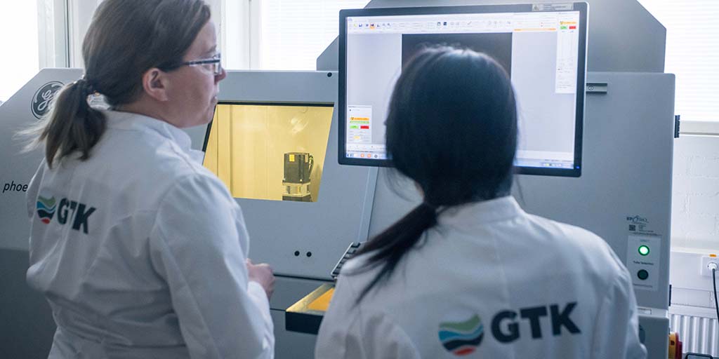
X-ray tomography is an imaging method that can be used to produce three-dimensional density maps on samples without destroying them. The method can be used for a wide selection of materials. In 2017, a GE Phoenix v|tome|x s 240 (XCT) X-ray tomography device was installed at GTK’s Espoo Research Laboratory. The device is especially designed for imaging geological and inorganic samples.
X-ray tomography is based on the absorption of X-rays by different materials. GTK’s device features the high-output 240 kV microfocus X-ray tube, which enables an accuracy of 5 micrometres. We also use the 180 kV nanofocus tube, which has a smaller output but enables an even better 1 micrometre accuracy. Samples to be imaged with the microfocus tube can be a maximum of 30 centimetres in diameter and 41 centimetres long, and weigh up to 10 kilos. When using the nanofocus tube, samples need to be significantly smaller. The best accuracy is achieved with samples under two millimetres in diameter.
Components in samples, such as mineral phases, must be identified with other methods, for example with SEM or EDS, from a two-dimensional surface. The information from the identification can then be applied to the density map.
GTK’s device also features a Deben’s sample holder assembly, which can be used to press or stretch the sample with a maximum force of 5 kN. At the same time, the sample can be cooled or heated to temperatures between -20 and +100 degrees Celsius.