Microanalysis laboratory
Microanalysis by means of an electron probe microanalyzer (EPMA) is an important mineralogical quantitative analysis method and often needed to supplement for example scanning electron microscopy (SEM) and energy dispersive spectrometry (EDS) as well as laser ablation-single collector-high resolution-inductively coupled plasma-mass spectrometry (LA-SC-HR-ICP-MS) analyses.
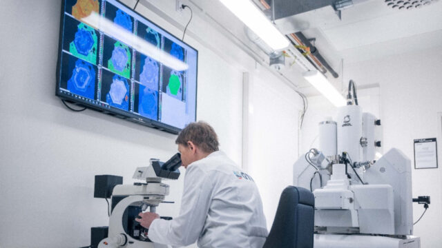
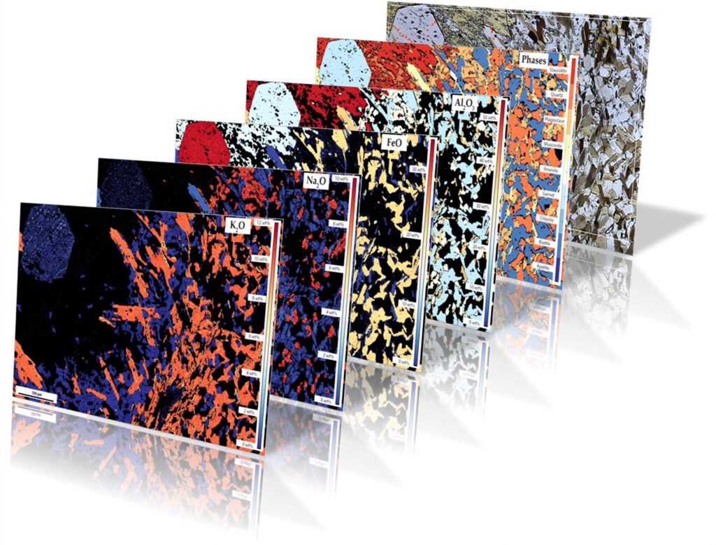
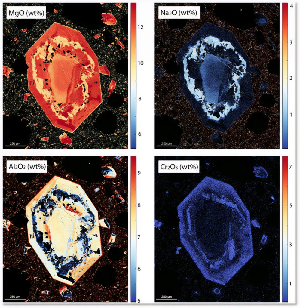
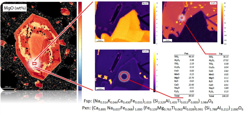
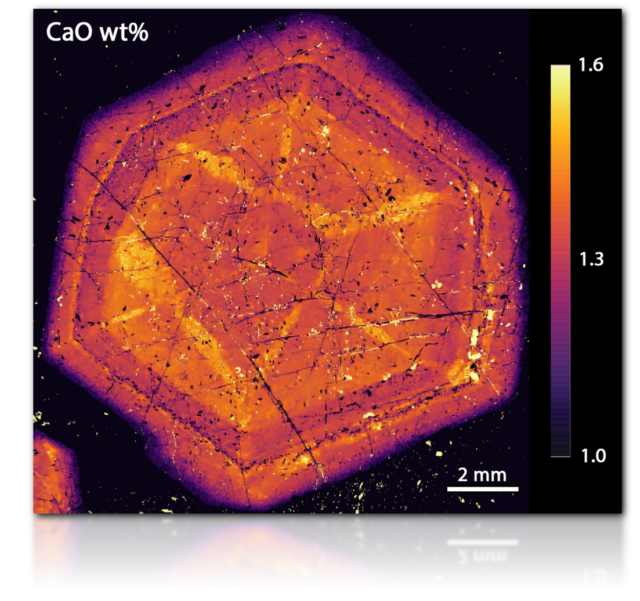
The GTK Research Laboratory in Espoo has recently (2021) acquired a JEOL JXA-iHP200F Hyper Probe field emission gun-EPMA (FEG-EPMA) that enables us to do quantitative analysis of nearly all elements on the periodic table (Be–U). In addition to a silicon drift detector (SDD)-EDS the state-of-the-art FEG-EPMA features five wavelength-dispersive spectrometers (WDS) used in routine quantitative analysis as well as high precision quantitative analysis even in sub-micron range and high accuracy quantitative element mapping on solid materials, such as natural minerals or synthetic compounds.
In routine analyses, the chemical-element-specific detection limits are around 0.01 wt.%, or as low as 10 ppm if a long measurement time is used for trace element analysis. The chemical elements to be analysed are selected beforehand. The SDD-EDS detector is used for qualitative analysis, together with high resolution back scattered and secondary electron imaging (BSE and SE) with a resolution down to 5 to 10 nanometers.
Point, profile, or grid analysis can be conducted with the smallest area in routine analysis of around 1–2 microns in diameter with an absolute elemental sensitivity around 10-15 g = 1 fg. Typically, mineral structures and other targets to be analysed must be identified with optical microscopy or SEM beforehand.
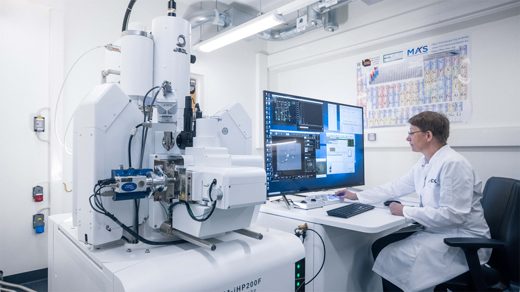
- Accelerating voltage up to 30 kV
- Beam current up to 3 µA
- Analyzed spot area Ø = <1 to 20 µm
- Information depth in the range of sub- to few microns
- Absolute elemental sensitivity around 10-15 g = 1 fg
- Detectable concentration range down to 10 ppm, typically 100s ppm
- Detectable elements range for atomic number Z ≥ 4 (Be) ≤ 92 (U)
- Quantification is standard reference based
- Sample preparation requires electrical conductivity by carbon coating and sample needs to have a flat polished surface
- Sample holder can hold up to 3 thin/thick sections or up to 6 one-inch epoxy mounts or up to 4 mounts with Ø = 3 cm
- Application software: JEOL SEM Center and JEOL PC-EPMA Ver 2 as well as the full Probe for EPMA software package
- Fully dry pumped vacuum system as well as sample insertion plasma cleaner to reduce hydrocarbon contamination
- 30 mm2 integrated UTW-SDD-EDS system with high sensitivity and an in-situ aperture wheel
Portfolio
Publications
2024
Astikainen O., Klemettinen L., Tammela J., Taskinen P., Michallik R.M., O’Brien H., Lindberg D., 2024. Industrial Deportment of Minor and Trace Elements in Direct Nickel Matte Smelting. JOM (TMS), https://doi.org/10.1007/s11837-024-06739-4
Liu S., Wang M., Gao Z., Liu X., Fan H-R., Butcher A., Lahaye Y., Michallik R., Jolis E., Lukkari S., 2024. The occurrence and distribution of Scandium resources in the world-class Bayan Obo REE-Nb-Fe deposit, China. European Geoscience Union General Assembly 2024 Abstract; doi.org/10.5194/egusphere-egu24-1809
Szentpéteri K., Cutts K., Glorie S., O’Brien H., Lukkari S., Michallik R.M., Butcher A., 2024. First in situ Lu–Hf garnet date for a lithium–caesium–tantalum (LCT) pegmatite from the Kietyönmäki Li deposit, Somero–Tammela pegmatite region, SW Finland. European Journal of Mineralogy 36, 433-448; doi.org/10.5194/ejm-36-433-2024
2023
Cutts K., Hölttä P., Lahtinen R., Kurhila M., Michallik R., 2023. U-Pb dating of garnet, monazite and apatite applied to understanding the polymetamorphic evolution of the Peräpohja and Kuusamo Belts, northern Finland. GOLDSCHMIDT 2023 Abstract; doi.org/10.7185/gold2023.17433
Sardisco L., Apeiranthitis N., Hirani J., Franzel M., Jolis E.M., Lukkari S., Michallik R.M., Tepsell J., PearceT.J., Butcher A.R., 2024. Battery Mineral Characterization—A Case Study of a Nickel Reference Material. Materials Proceedings 2023, 15(1), 83; doi.org/10.3390/materproc2023015083
